Physical Address
304 North Cardinal St.
Dorchester Center, MA 02124
Physical Address
304 North Cardinal St.
Dorchester Center, MA 02124
Rewrite the content and keep the original meaning and format, and add 5 more H2, the content is “
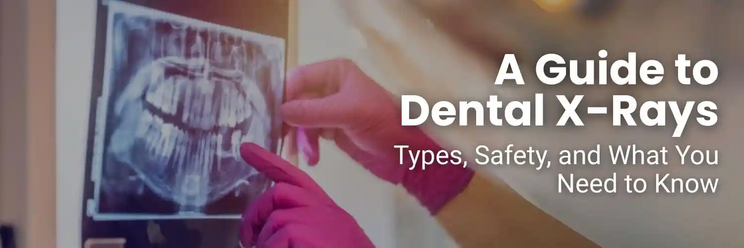
Your dentist can spot visible plaque and chips, but the real story of your oral health lies beneath the surface. Dental radiographs expose hidden decay between tight contacts, pinpoint early bone loss around implants, verify root-canal fillings, and even reveal cysts or unerupted teeth—problems that a mirror alone will never catch. Because timely treatment depends on seeing the unseen, dental X-rays are as indispensable to modern dentistry as the stethoscope is to medicine.
Today’s imaging is faster and far safer than the film era. High-frequency, handheld generators such as the Genoray Port X III Portable X-Ray and the Runyes Handheld Portable X-Ray pair with digital sensors to slash exposure times while giving dentists the flexibility to capture crisp images chairside—ideal for children, anxious patients, and wheelchair users. These advances, coupled with lead aprons, thyroid collars, and tight beam collimation, keep doses well below everyday background radiation, reinforcing best-practice radiation safety standards like ALARA (As Low As Reasonably Achievable).
In the sections that follow, we’ll break down the main types of dental X-rays—from bitewings and periapicals to panoramic “panos” and 3-D CBCT scans—explain when each view is ordered, and compare traditional vs. digital dental X-rays for dose and clarity. We’ll wrap up with patient FAQs on frequency, safety during pregnancy, and insurance coverage, giving you the knowledge to discuss your imaging plan confidently at your next appointment.

|
Category |
How It’s Taken |
Best-Use Scenarios |
|
Intraoral X-Ray • Checking root length, periapical infections, or post-root-canal fills (periapicals) •Mapping erupting teeth of supernumeraries on a single arch (occlusal views) |
A small digital sensor or film is placed inside the mouth while the X-ray head rests just outside the cheek. |
• Detecting cavities between teeth (bitewings) |
|
Extraoral Dental Imaging • Evaluating jaw joints, fractures, or orthodontic growth patterns (cephalogram) • Producing 3D data for guided surgery and airway studies (CBCT) |
The detector stays outside the mouth; the beam traverses the jaws from an external vantage point. |
• Capturing the entire upper and lower jaws in one shot for wisdom-tooth or implant planning (panoramic X-ray) |
Key Takeaway
Together, these three intraoral views deliver a comprehensive, low-dose toolkit that lets dentists diagnose everything from tiny interproximal lesions to deep-root pathologies—without leaving the exam chair.
Panoramic X-Ray (OPG)
3-D Cone-Beam CT (CBCT)
Cephalometric Scan
Together, panoramic, CBCT, and cephalometric imaging provide a full-field view that complements intraoral details—equipping dentists with a complete map before they drill, extract, or move a single tooth.
Digital vs. Film
Switching from traditional D-speed film to modern CMOS or PSP sensors slashes exposure by 60–80 %. Digital plates are 10-20× more sensitive to X-ray photons, so the generator can operate at shorter pulses and lower milliampere settings while still delivering razor-sharp images.
|
Metric |
Film (D-speed) |
Digital Sensor |
|
Typical bitewing dose* |
170 µSv |
30–60 µSv |
|
Retake rate |
Higher (processing errors) |
Lower (instant preview) |
|
Environmental impact |
Chemical fixer waste |
None |
*Micro-sievert values vary by unit and technique.
Four Layers of Protection
Combined with digital capture, these safeguards make dental radiography one of the lowest-dose modalities in medical imaging—providing critical diagnostic data while keeping patient exposure comfortably within international safety guidelines.
From the pinpoint detail of a bitewing to the full-arch sweep of a panoramic or 3-D clarity of a CBCT, every X-ray view has a specific job: detect hidden decay, gauge bone, map nerves, or plan surgery. Knowing which image answers which clinical question lets you ask smarter questions and give truly informed consent—confident that each exposure has a clear payoff.
Thanks to high-sensitivity digital sensors, tailored beam collimation, and ALARA protocols, today’s dental imaging delivers vital data at fractions of the radiation once required. But frequency still matters. Caries-prone kids may need bitewings twice a year; low-risk adults might stretch to every two or three. A panoramic might be once in five years unless wisdom-tooth or implant planning calls for it, and a CBCT only when three-dimensional data will change the outcome.
Talk with your dentist about your medical history, cavity risk, and upcoming treatments to craft an imaging schedule that’s personalized, safe, and cost-effective. When you understand the purpose and protection behind each dental X-ray, you can partner confidently in your care—securing the clearest picture of oral health while keeping exposure as low as science allows.
”