Physical Address
304 North Cardinal St.
Dorchester Center, MA 02124
Physical Address
304 North Cardinal St.
Dorchester Center, MA 02124
Rewrite the content and keep the original meaning and format, and add 5 more H2, the content is “
Dental imaging has come a long way from traditional 2D X-rays to advanced 3D imaging, significantly improving diagnostic accuracy and treatment planning. While 2D radiographs have been a standard tool for years, they often come with limitations in depth perception, anatomical distortion, and incomplete visualization. Dentists using only 2D imaging may struggle to detect hidden infections, bone loss, or precise nerve positioning, leading to less predictable treatment outcomes.
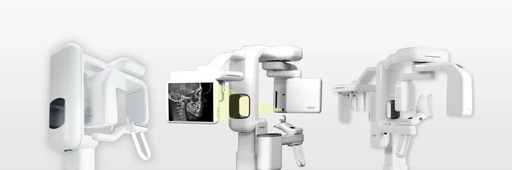
The introduction of 3D imaging, particularly Cone Beam Computed Tomography (CBCT), has revolutionized dental diagnostics. Unlike conventional X-rays, CBCT provides a detailed 360-degree view of the patient’s teeth, bone structure, nerve pathways, and soft tissues, allowing for highly precise and patient-specific treatment planning. This advancement is especially crucial in implantology, orthodontics, endodontics, and oral surgery, where precision is key to success.
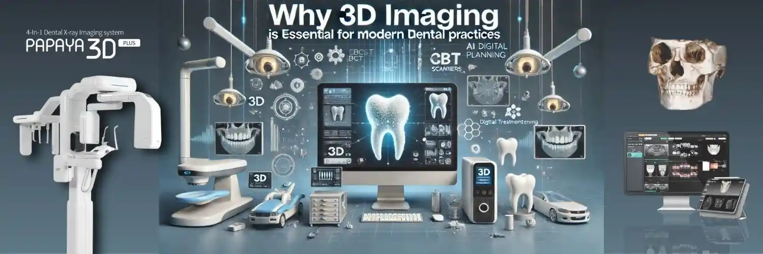
One of the most cutting-edge CBCT systems available today is the Genoray Papaya 3D CBCT. It offers high-resolution imaging with minimal radiation exposure, making it a safer and more effective option for both dentists and patients. The system’s versatility in capturing detailed 3D scans for various dental procedures ensures that clinicians have a complete picture before proceeding with any treatment.
This blog will explore why 3D imaging is now an essential tool in modern dentistry, focusing on its advantages in diagnostics, treatment planning, and overall patient care. As dental technology advances, embracing 3D imaging solutions like the Genoray Papaya 3D CBCT will be crucial for clinics looking to provide accurate, efficient, and high-quality dental care.
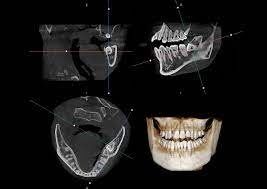
Achieving highly accurate diagnostics is crucial in dentistry, and Cone Beam Computed Tomography (CBCT) has become an indispensable tool for detailed visualization and precise treatment planning. Unlike traditional 2D X-rays, which offer only flat, limited views of dental structures, CBCT imaging captures high-resolution, three-dimensional scans, allowing dentists to assess bone quality, nerve pathways, and hidden dental issues with unparalleled clarity.
One of the standout CBCT systems, the Genoray Papaya 3D CBCT, enhances diagnostic precision by providing clear, distortion-free 3D images with minimal radiation exposure. This advanced imaging system enables dentists to detect cysts, impacted teeth, periapical infections, and even early signs of bone loss, which might go unnoticed with conventional imaging techniques.
Accurate visualization of bone density and nerve positioning is particularly critical for dental implant placement, orthodontics, and complex root canal treatments. The Genoray Papaya 3D CBCT allows dentists to measure anatomical structures with millimeter precision, ensuring flawless implant positioning, reduced surgical risks, and improved treatment outcomes.
By incorporating high-resolution 3D imaging into clinical workflows, dental professionals can prevent treatment failures, enhance patient safety, and improve long-term oral health results. With CBCT technology like the Genoray Papaya 3D CBCT, dentists can make data-driven decisions, ensuring minimally invasive, highly successful treatments for their patients.
In modern dentistry, precision and predictability are critical for successful treatment outcomes, especially in implantology, orthodontics, and endodontics. 3D imaging technology, particularly Cone Beam Computed Tomography (CBCT), has transformed treatment planning by offering detailed anatomical visualization that traditional 2D X-rays cannot provide.
For dental implant placement, precise knowledge of bone density, nerve positioning, and sinus location is essential to avoid complications. The Genoray Papaya 3D CBCT provides high-resolution 3D scans, allowing dentists to plan implant placements with millimeter accuracy, reducing the risk of nerve damage or improper positioning. This leads to higher success rates and long-term stability of implants.
In orthodontics, 3D imaging helps in assessing jaw alignment, tooth positioning, and airway analysis, enabling orthodontists to design personalized treatment plans. The clear, full-view scans from the Genoray Papaya 3D CBCT ensure better bracket placement, accurate tooth movement predictions, and reduced treatment time.
For endodontic procedures, detecting hidden canals, root fractures, or periapical infections is often challenging with conventional X-rays. CBCT imaging enhances diagnostic accuracy, ensuring precise root canal treatment with fewer complications.
By utilizing advanced 3D imaging like the Genoray Papaya 3D CBCT, dentists can create customized, patient-specific treatment plans, leading to safer, more efficient procedures and improved patient satisfaction. This technology ensures that complex treatments are executed with confidence, accuracy, and long-term success.
One of the biggest advantages of 3D imaging in modern dentistry is its ability to make treatments minimally invasive and safer for patients. Traditional methods often require exploratory surgery or multiple X-rays to assess bone structure, nerve pathways, or hidden dental issues. However, 3D Cone Beam Computed Tomography (CBCT) eliminates the need for unnecessary surgical intervention by providing detailed, real-time visualization of oral and maxillofacial structures before treatment begins.
Compared to medical CT scans, CBCT imaging offers significantly lower radiation exposure while delivering high-resolution, 360-degree views of the teeth, bone, and surrounding tissues. This ensures that dentists can accurately plan implant placements, extractions, and root canal treatments without subjecting patients to excessive radiation. The ability to pinpoint anatomical structures precisely reduces procedural risks and enhances treatment predictability.
With 3D imaging, dentists can perform more precise and targeted treatments, leading to faster recovery times, reduced trauma, and minimal post-operative discomfort. In implantology, orthodontics, and oral surgery, the use of CBCT imaging ensures optimal outcomes with the least invasive approach possible.
By integrating 3D imaging technology into their practice, dentists can enhance patient safety, improve surgical precision, and minimize treatment complications, making procedures safer, more comfortable, and highly effective for long-term success.

The integration of 3D imaging with CAD/CAM and digital dentistry has significantly improved workflow efficiency and patient communication in modern dental practices. By eliminating the delays associated with traditional impression-taking, lab-dependent restorations, and manual treatment planning, Cone Beam Computed Tomography (CBCT) enhances precision, speed, and case predictability.
3D imaging seamlessly integrates with CAD/CAM systems, allowing dentists to digitally design crowns, bridges, and implants with pinpoint accuracy. By generating precise 3D models of the patient’s anatomy, CBCT technology ensures better-fitting restorations and improved prosthetic designs. This reduces treatment time, lab turnaround delays, and manual errors, creating a smoother workflow for dentists and patients alike.
Another significant advantage of 3D imaging is its impact on patient communication and treatment acceptance. With high-resolution 3D visuals and AI-driven simulations, dentists can educate patients about their oral condition and proposed treatment plan in real-time. Seeing clear, interactive visuals helps patients better understand their diagnosis, increasing trust and confidence in the recommended procedures.
This enhanced communication also leads to faster treatment approvals, as patients feel more informed and reassured about their choices. As a result, case acceptance rates improve, and clinics can deliver high-quality treatments efficiently and effectively.
By incorporating 3D imaging into their digital workflows, dentists can optimize operations, reduce chair time, and create a more engaging, transparent, and patient-friendly experience in their practice.
The adoption of 3D imaging in dentistry has transformed diagnostic accuracy, treatment planning, and patient care, making it an essential tool for modern dental practices. Unlike traditional 2D X-rays, Cone Beam Computed Tomography (CBCT) provides detailed 3D visualization of teeth, bone structure, and nerve positioning, ensuring greater precision in complex procedures such as implantology, orthodontics, and endodontics.
With enhanced efficiency, minimized surgical risks, and improved patient communication, 3D imaging enables faster diagnoses, predictable treatment outcomes, and a seamless digital workflow. Technologies like Genoray Papaya 3D CBCT offer high-resolution imaging with low radiation exposure, making diagnostics safer, faster, and more effective.
To stay ahead in modern dentistry, clinics must embrace 3D imaging to improve workflow efficiency and patient confidence. Investing in CBCT technology not only enhances treatment success rates but also helps dentists deliver superior care with minimal invasiveness.
As digital dentistry continues to evolve, 3D imaging is no longer an option—it is a necessity for clinics striving for precision, efficiency, and long-term patient trust. Now is the time for dentists to integrate 3D imaging into their practice and elevate the standard of dental care.
”