Physical Address
304 North Cardinal St.
Dorchester Center, MA 02124
Physical Address
304 North Cardinal St.
Dorchester Center, MA 02124
Rewrite the content and keep the original meaning and format, and add 5 more H2, the content is “
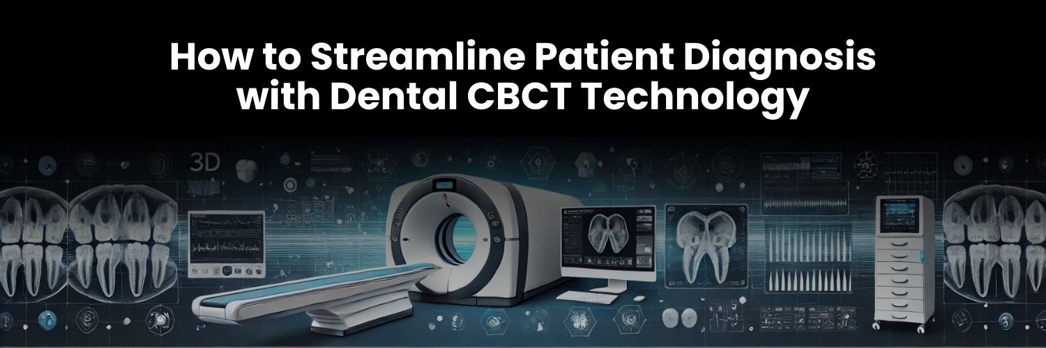
Accurate diagnosis is the foundation of effective dental treatment. Whether planning an implant, performing a root canal, or assessing jaw alignment, precise imaging is essential for achieving successful outcomes. Traditional 2D X-rays, while useful, have limitations in providing a comprehensive view of complex dental structures. This is where Cone Beam Computed Tomography (CBCT) has revolutionized modern dentistry, offering detailed 3D imaging that enhances diagnostic accuracy and treatment precision.
The Genoray Papaya 3D Plus CBCT is one of the most advanced imaging solutions available today. It provides high-resolution, full 3D scans of the oral and maxillofacial region, allowing dentists to evaluate bone structure, nerve positioning, and anatomical variations with exceptional clarity. Unlike conventional imaging, CBCT offers multi-angle visualization, helping clinicians identify hidden issues such as impacted teeth, fractures, and periapical lesions that may go unnoticed on 2D radiographs.
Beyond accuracy, CBCT technology significantly improves efficiency and workflow integration. It reduces the need for multiple scans, minimizes exploratory procedures, and enhances patient communication by allowing practitioners to visually explain diagnoses and treatment plans. With quick scan times and low radiation exposure, CBCT provides a safe, patient-friendly imaging experience.
As dental technology advances, adopting CBCT systems like the Genoray Papaya 3D Plus is no longer optional but essential. It enables precise treatment planning, improved patient outcomes, and a more streamlined diagnostic process, positioning dental practices at the forefront of modern, high-quality care.

Cone Beam Computed Tomography (CBCT) has transformed dental imaging by providing detailed 3D visualization, offering a significant advantage over traditional 2D X-rays. Unlike conventional radiographs, which capture flat images with limited depth perception, CBCT produces high-resolution, three-dimensional scans of the oral and maxillofacial region. This allows dentists to view anatomical structures from multiple angles, leading to more precise diagnoses and treatment planning.
One of the biggest benefits of CBCT is its higher diagnostic accuracy, especially in complex cases. It enables clear identification of root fractures, impacted teeth, bone density variations, sinus conditions, and nerve positioning—all of which are crucial for procedures like implant placement, endodontic treatments, and orthodontic planning. By eliminating the guesswork associated with 2D imaging, CBCT allows for more predictable and successful outcomes.
CBCT technology also enhances patient safety and procedural efficiency by reducing the need for exploratory surgeries. In cases where traditional imaging may not reveal the full extent of an issue, dentists would typically rely on invasive methods to assess the problem. CBCT minimizes this need by providing precise anatomical insights upfront, helping practitioners make well-informed clinical decisions while reducing unnecessary procedures and patient discomfort.
With its superior imaging capabilities, CBCT enhances diagnosis, improves treatment accuracy, and increases patient confidence—making it an essential tool for modern dental practices aiming to deliver high-quality, minimally invasive care.

Cone Beam Computed Tomography (CBCT) has become an indispensable tool in modern dentistry, providing precise 3D imaging that enhances diagnostics and treatment planning. Its ability to capture detailed anatomical structures makes it particularly valuable in several key areas of dentistry.
Implant Planning
CBCT is crucial for dental implant placement, as it allows dentists to accurately analyze bone density, volume, and nerve positioning before surgery. This helps determine whether bone grafting is needed and ensures implants are placed at the optimal angle and depth, reducing complications and improving long-term success rates.
Endodontics
For root canal treatments, CBCT imaging aids in detecting root fractures, extra canals, and periapical lesions that may not be visible on 2D X-rays. It provides a clearer view of root morphology, allowing endodontists to perform more precise and effective treatments, reducing the risk of failed procedures.
Orthodontics
CBCT is widely used in orthodontic treatment planning, helping assess jaw alignment, tooth movement, and airway conditions. It allows orthodontists to evaluate skeletal discrepancies, impacted teeth, and potential obstructions, leading to more accurate and customized treatment plans.
Oral Surgery
For extractions, sinus lifts, and bone grafting, CBCT provides a detailed map of anatomical structures, reducing surgical risks. It helps identify nerve pathways, sinus cavities, and bone abnormalities, ensuring safer and more efficient surgical procedures.
By offering unparalleled accuracy and detail, CBCT technology significantly enhances treatment planning across multiple dental specialties, making it a must-have tool for modern dental practices.
Cone Beam Computed Tomography (CBCT) has revolutionized dental workflows by significantly improving diagnostic speed, treatment planning accuracy, and overall efficiency. Unlike traditional imaging methods, which often require multiple scans and interpretation delays, CBCT provides instant, high-resolution 3D images, enabling faster and more precise diagnoses. This reduces the time spent on treatment planning and patient consultations, allowing for quicker decision-making and more effective treatments.
One of CBCT’s biggest advantages is its seamless integration with digital workflows, including CAD/CAM systems, AI-based treatment planning tools, and 3D printing technologies. With CBCT-generated images, dentists can create digital impressions, design custom prosthetics, and simulate surgical procedures with greater accuracy. This not only improves procedural outcomes but also enhances communication between specialists, dental labs, and patients.
By offering detailed pre-treatment insights, CBCT reduces the need for unnecessary exploratory procedures and multiple patient visits. For example, in implantology, dentists can use CBCT data to virtually plan implant placement before surgery, reducing chair time and optimizing implant success. Similarly, in orthodontics, CBCT aids in precise tooth movement analysis, minimizing adjustments and treatment duration.
With its ability to streamline workflows, improve accuracy, and enhance patient experiences, CBCT has become an essential tool for modern dental practices. Dentists who adopt CBCT technology benefit from faster case turnaround times, improved efficiency, and higher patient satisfaction, making their practice more competitive in today’s digital-driven dental industry.

One of the key advantages of Cone Beam Computed Tomography (CBCT) is its patient-friendly design, offering a safer, faster, and more comfortable imaging experience compared to traditional methods. Unlike conventional CT scans, which expose patients to high levels of radiation, CBCT operates with significantly lower radiation doses while still providing highly detailed 3D images. This makes it a safer option for routine dental diagnostics, particularly for children and patients requiring multiple scans.
CBCT also enhances patient confidence by allowing dentists to visually explain diagnoses with clear, three-dimensional images. Patients can see detailed views of their bone structure, nerve pathways, and dental conditions, making it easier for them to understand their treatment options. This transparency fosters trust and engagement, leading to better patient compliance and satisfaction.
Another major benefit of CBCT technology is that it is completely pain-free, quick, and non-invasive. The scanning process takes only a few seconds, eliminating the discomfort associated with traditional impression-taking or multiple 2D X-rays. Patients simply stand or sit while the scanner rotates around their head, making the procedure stress-free.
With lower radiation exposure, enhanced diagnostic clarity, and a comfortable scanning process, CBCT improves both patient safety and overall experience. By adopting CBCT technology, dental clinics can provide high-quality, patient-centered care, ensuring more accurate diagnoses while prioritizing patient well-being.
Cone Beam Computed Tomography (CBCT) has transformed dental diagnostics by providing unmatched precision, efficiency, and patient comfort. Its ability to generate high-resolution 3D images streamlines diagnosis and treatment planning, reducing the need for multiple scans and exploratory procedures. By enhancing accuracy in implantology, endodontics, orthodontics, and oral surgery, CBCT ensures better clinical outcomes and improved patient safety.
For dentists, investing in CBCT technology means greater workflow efficiency, reduced chair time, and a more seamless integration with digital tools like CAD/CAM and AI-driven treatment planning. This not only optimizes treatment procedures but also enhances patient confidence through clearer communication and visualization of diagnoses.
Adopting CBCT technology positions a dental practice at the forefront of modern, high-quality dentistry, helping practitioners stay competitive in an evolving industry. With its ability to deliver faster, safer, and more accurate diagnoses, CBCT is no longer just an innovation—it is a necessity for dentists committed to providing top-tier, patient-centric care. Investing in CBCT today means ensuring a future-ready practice that stands out in precision, efficiency, and excellence.
”